Diagnostic exams
The Bogota Laser Diagnostic Center is equipped with state-of-the-art ophthalmic analysis technology, enabling a thorough examination of the eye and facilitating the identification of the most beneficial course of action for each patient. This advanced technology aids in accurately diagnosing eye conditions and determining the appropriate treatment plan.
Elevation topography
We employ advanced ophthalmic analysis technology to thoroughly assess the cornea, including its anterior and posterior surfaces, as well as the anterior chamber and iris of the eye. This comprehensive evaluation is particularly essential for cases involving conditions like keratoconus, where eye surgery or intra-stromal rings may be required. It also serves as a reliable indicator of the risk of future complications with laser surgery, such as ectasias. Moreover, this examination provides a more precise calculation of the intraocular lens in individuals with a history of refractive surgery. We utilize cutting-edge equipment, such as the Pentacam HR by Oculus and Sirius by Schwind, to perform these assessments.
Recommendations: If you wear soft contact lenses, remove them at least 3 days before the examination; if you wear hard contact lenses, remove them 8 days in advance. No dilation is required.
Diagnostic Center: Pentacam, SiriusPachymetry
One of the essential aspects of our diagnostic process is measuring the thickness of the cornea in microns. This measurement allows us to determine the most suitable and safest procedure for correcting visual defects, including astigmatism, hyperopia, myopia, presbyopia, and keratoconus. To perform this evaluation, we utilize advanced equipment such as the Pentacam HR by Oculus or Sirius by Schwind, specifically designed for pachymetry.
Recommendations: If you wear soft contact lenses, remove them at least 3 days before the examination; if you wear hard contact lenses, remove them 8 days in advance. No dilation is required.
Diagnostic Center: Pentacam, Sirius
Aberrometry
This examination uses laser light rays to accurately diagnose visual defects without the subjectivity of subjective refraction in optometry. It measures the present refractive error and other optical defects called aberrations, which are converted into measurements through mathematical formulas to determine the appropriate treatment with the excimer laser. This system is known as a personalized treatment of visual defects. At Bogota Laser Ocular Surgery Center, we have three types of equipment to perform this examination, depending on the laser that will be used for the patient and the procedure defined by the ophthalmologist. These are Peramis, Idesing, and Wavefront.
Recommendations: If you wear soft contact lenses, remove them at least 3 days before the examination; if you wear hard contact lenses, remove them 8 days in advance. No dilation is required.
Diagnostic Center: Wavefront, I desing, Peramis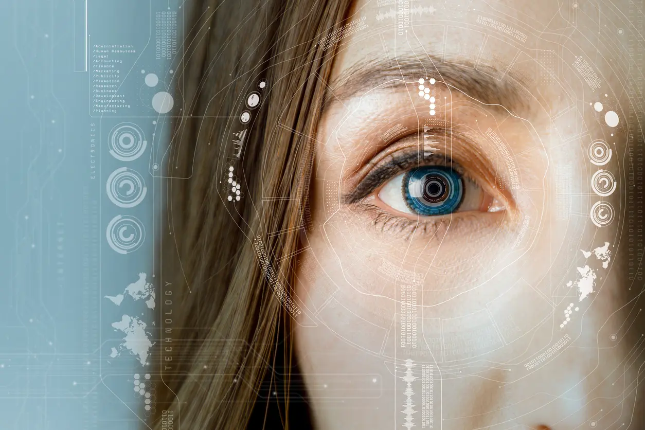
Pupillometry
Recommendations: If you wear soft contact lenses, remove them at least 3 days before the examination; if you wear hard contact lenses, remove them 8 days in advance. No dilation is required.
Diagnostic Center: Sirius,Peramis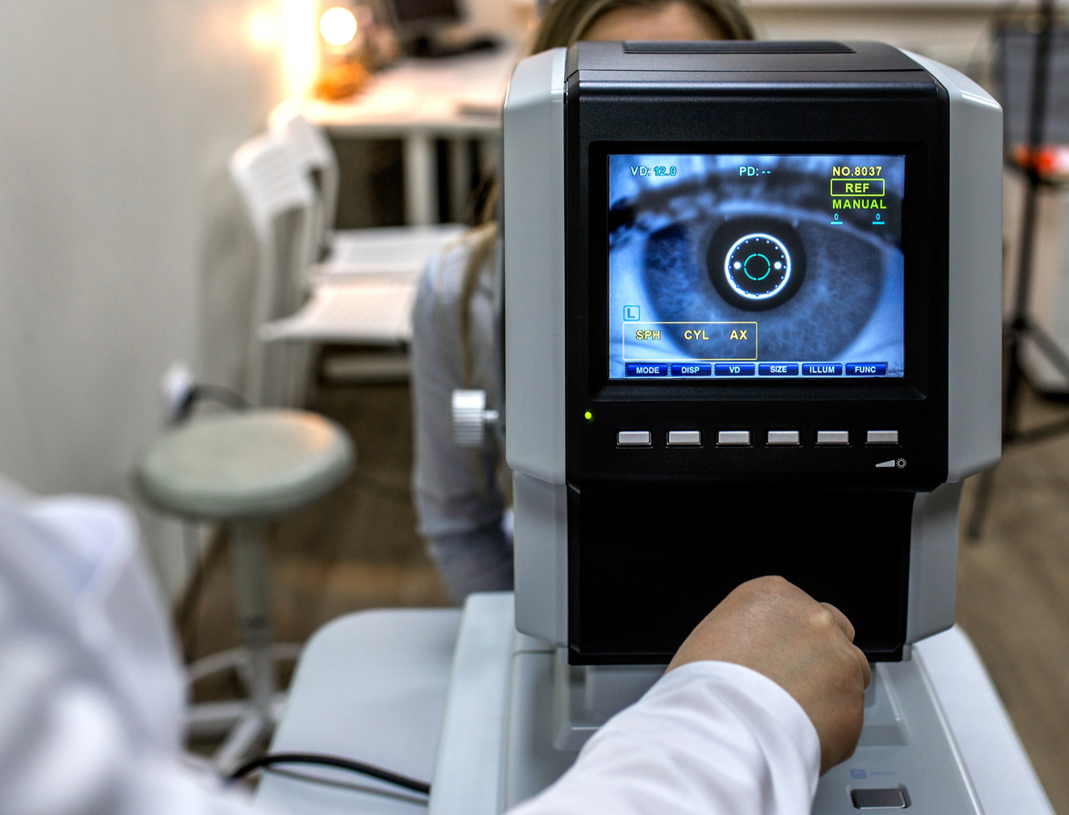
Autorefraction
It provides a comprehensive analysis of visual defects (astigmatism, hyperopia, myopia, presbyopia, and keratoconus) under different pupil sizes, as well as the accompanying optical aberrations.
Recommendations: If you wear soft contact lenses, remove them at least 3 days before the examination; if you wear hard contact lenses, remove them 8 days in advance. No dilation is required.Diagnostic Center: Autorefractómetro, Nidek, Autorefractor Visuref 100
Optical coherence tomography (oct)
Recommendations: Corneal OCT and anterior chamber angle measurement do not require dilation. For macular and optic nerve OCT, please take precautions for your travel arrangements, ensure you have assistance, or do not drive. This examination requires reading, and the results will be available within 3 to 5 business days after the examination.
Diagnostic Center: Optovue OCT
Ocular biometry
Recommendations: No specific preparation is required. In some cases, anesthesia may be applied, but only if ultrasound is necessary.
Diagnostic Center: Lenstar Biometer, Contact Biometer
Dry eye analysis – keratograph
The Keratograph 5M enables various studies to be conducted to evaluate different aspects of tear function and ocular health. These include tear meniscus height, tear film sensitivity and stability, lipid layer thickness, tear film dynamics, meibography test, and bulbar redness recording (hyperemia).
Recommendations: If you wear contact lenses, remove them before the examination, and avoid using any medication or artificial tears 2 hours before the test to avoid affecting the results.
Endothelial cell count
The specular microscopy examination is the only analysis available for assessing corneal vitality and allows for determining and calculating the cornea's survival time without damaging its structure. The examination is performed with the Iconon Microscope Specular.
Recommendations: No dilation is required.
Air tomography ora
Verion – Bogotá Láser
Contrast vision
ORA – Bogotá Láser
Our technology brands
 .
.